Breast cancer screening - what to expect?
About Breast Cancer Screening
Women with an increased risk of developing breast cancer can be seen at the NKI Center for Early Diagnostics screening clinic. Each case is discussed in a multidisciplinary team meeting (MDO) prior to the periodic check-up. All specialists involved in the screening clinic are present at this meeting.
At the Screening Clinic, you will receive comprehensive information and advice tailored to your personal situation. We also conduct periodic breast cancer screenings. Depending on your situation, this may include a mammogram or MRI scan of the breasts.
Your nurse specialist will discuss the results of the mammogram and/or MRI with you at your next appointment, which will usually be about a week later.
In some cases, additional examinations are needed, such as an ultrasound, liquid biopsy, or tissue biopsy.
Learn more about our tests:

What is an MRI Scan?
An MRI scan provides detailed images of breast tissue. It uses radio waves in combination with a large, powerful magnet to generate signals in the body, which are then captured by a computer using an antenna, and processed into an image. This scan does not use X-rays. The MRI scan visually slices the breasts into thin sections. You will lie down on a bed that can slide into a tunnel for the scan. The MRI machine can be quite loud, so you will receive headphones with music to help block out the sounds. A contrast agent will be administered through an IV, which may cause a warm sensation in your body, a dry mouth, and a need to urinate.
The MRI scan will take about 25 minutes.
What is an Ultrasound?
Ultrasound is a technique used to visualize a body part using sound waves. The device emits inaudible sound waves that bounce back within your body, creating an image on the monitor. The radiologist applies a gel to the skin at the area currently being examined, and moves the device over the skin. You can usually watch the process on the monitor. If the radiologist detects any abnormalities, they may ask you whether they can perform a liquid or tissue biopsy immediately.
An ultrasound will take about 15 minutes.
What is Mammography?
A mammogram is an X-ray image of the breasts.
How is a Mammogram Performed?
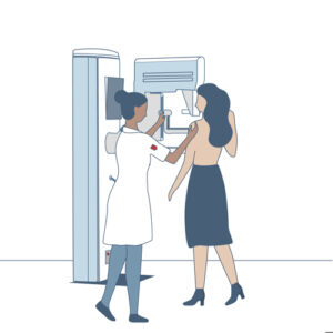
You will be asked to remove your upper clothing in a special dressing room adjacent to the examination area. Then, you will be guided into the examination room by the technician.
The breast is placed between two plates that compress it. This spreads out the breast tissue, revealing any abnormalities on the image.
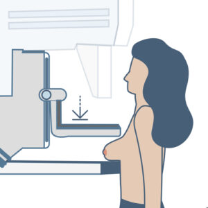
The technician will take at least two images of each breast: one from above and one from the side. Sometimes we will take additional images.
Many women find the compression uncomfortable or painful. The pressure lasts only a few seconds. If you experience pain during the procedure, please inform the technician, who will try to accommodate you as much as possible.
A mammogram will take about 15 minutes.
What is a liquid biopsy?
During a liquid biopsy, the radiologist uses a thin needle to extract cells or fluid from the body. The exact location for the liquid buiopsy is usually determined with the help of an ultrasound. The extracted material is then examined in the laboratory. A pathologist will determine whether the findings are benign or malignant.
A liquid biopsy will take about 15-30 minutes.
After the liquid biopsy
The results of the biopsy will usually be available within an hour. Your nurse practitioner will discuss the results with you.
What is a tissue biopsy?
A tissue biopsy is a procedure in which a small piece of tissue is removed. This piece of tissue is called a sample. The tissue biopsy is generally performed under local anesthesia. The tissue sample will be taken through a hollow needle. To determine the precise location for the biopsy, we often use an ultrasound or a CT scan. A pathologist examines the biopsy sample in the laboratory to determine whether the abnormality is benign or malignant. Additionally, the pathologist can identify specific characteristics of any tumor, which helps us tailor a potential treatment to your situation.
A tissue biopsy will take about 15-45 minutes.
After the Biopsy
The results of a biopsy will generally be available after five weekdays. Your nurse practitioner will discuss the results with you.
What is a stereotactic biopsy?
In a stereotactic biopsy, small pieces of tissue (tissue samples) are taken from an abnormality in the breast. This procedure uses X-ray imaging to be able to locate the abnormality more precisely. The tissue sample will be analyzed at the laboratory to determine whether there are malignant cells. The stereotactic biopsy is performed in the main building of the Netherlands Cancer Institute.
A stereotactic biopsy will take about one hour.
After the stereotactic biopsy
The results of the biopsy will generally be available after five working days. Your nurse practitioner will discuss the results with you.